

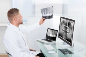
Dental sensors are the current modern technology that has improved the dental imaging industry. They have brought efficiency to radiography departments and comfort to the patients. Dental x-ray sensors technology uses digital x-ray sensors connected to application software. The sensors work with intraoral cameras that provide x-ray images for clinicians and dentists helping in diagnosing teeth and other oral complications.
The digital dental x-ray sensors and the intraoral cameras minimize exposure to radiation, and they are sized to accommodate both children and adult mouths. This new technology is shaping when Starting a Dental Practice because of its accurate, more detailed, and high-quality images it produces. It takes a short time to get results, and it offers a level of comfort for patients.
How do digital sensors work?
The dental x-ray sensors technology works in that the sensor is connected to an interface that is either mounted on a wall or a counter. The interface is then attached using a USB cable to your computer. Digital x-ray sensors have cameras. Hence, they take the images and relay them to the interface, and with the use of the software, the images are displayed on the computer.
Advantages of Dental Digital Imaging Technology
Digital dental sensors are a critical component for a busy Dental Practice. Understanding some of the key components involved in implementing a new sensor in your office will help your team make sure there are no surprises.
- Digital Imaging X-Ray reveals more by detecting small hidden points of decay in between the teeth, bone infections, abscesses or cysts, gum disease, tumors, and abnormalities that visual dental examination cannot detect.
- Digital imaging is instant. That is to say that it can be viewed on the computer screen and help in enhancing detail and contrast, giving clarity to specialists who get results transmitted electronically to them without losing the quality.
- Digital imaging help in giving results quickly hence enabling early detection and treatment, which saves time, money, and discomfort the patient may be feeling.
- It provides an eco-friendly environment because it eliminates the disposal of lead foil and other hazardous wastes from chemical processing.
- Digital imaging uses micro-storage technology that enables more excellent data storage on small drives that saves space.
- Digital radiographs can be shared with other dentists via sending them to compatible computer technology or by printing a photo to dentists who do not have the compatible technology.
- Digital sensors need 50%-80% less radiation compared to film. This is because technology is more sensitive to x radiation.
- Digital imaging offers secure storage to electronic patient records that can be sent to insurance companies for payment, helping reduce treatment disruption.
- The technology features such as colorizing, 3-D, flip, sharpness, and zoom assist in the detection and interpretation that help in diagnosis and treatment.
Top dental x-ray sensors in the market.
Dental sensors, even those of the same brand, vary significantly in their quality. It is thus valuable to establish action levels for the acceptance of newly delivered sensors and to use objective image quality control for commissioning purposes and periodic checks to ensure the high performance of individual digital sensors. Most current dental sensors generally perform well in terms of spatial and contrast resolutions, and in terms of exposure latitude.
Here is a comparison of different dental x-ray sensors used in dentistry.
1. Schick 33 ($3,490.00)
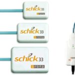
Schick 33 gives a leading image quality, which provides an improvement to diagnosis. It has expansive system choices, amazing capabilities in image enhancement, and top service from the support team. It offers an unprecedented resolution for detailed images. Its sensors use replaceable cables that are available in six feet, nine feet, and a shorter version of three feet.
The sensor cables are coated with silicon and are Kevlar reinforced to keep it running. Its interface really gives you a durable USB connection. The Schick 33 has multiple options to fit according to your treatment rooms. It has 3 sensor sizes, its cables are of many lengths, it has various enhancement capabilities to suit the patient’s needs and the specialist personal preferences.
Clinicians can entirely depend on Schick 33 superior image quality for featuring crisp lamina dura, bold bone trabeculation and giving a clear dental enamel junction. The intraoral sensor provides a leading theoretical resolution and brings a level of clarity, detail, and sharpness to digital radiography. Image clarity is a critical aspect of detecting defects, caries, and diseases.
Schick 33 can be customized to fit into your practice preference on digital imaging. It is said to be your own system, your own terms. Users benefit from good images, and through the replaceable DIY cables, it gives such a long-serving time. Schick has gone to another level in service because their systems are installed, supported, and serviced by Schick’s highly qualified engineers.
2. RVG 6200 ($3,100.00)
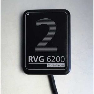
The RVG 6200 dental x-ray sensors have exceptional image quality with a 24lp/mm image resolution. It has customizable views with a built-in perio and dentin-enamel junction filters. It gives a simple, comfortable position with its secure acquisition process position and a thinner back cable.
It has high-quality image customization, logical caries detection software, which is optional, high imaging flexibility, and intuitive maintenance and installation. Its image processing software makes accurate and efficient diagnoses.
The RVG 6200 sensor head has a sleek design with rounded corners that create excellent comfort for both the patient and the dentist. The cable is thin and streamlined and more flexible for patient satisfaction. It has a complete set of devices for positioning, which achieves a higher degree of accuracy, faster image acquisition with consistent images.
The image acquisition of the RVG 6200 is fast and straightforward with the only position, expose, then view. Its twain compatibility entails that the sensor can be linked with much dental practice management and imaging software.
It has an easy installation, and its maintenance process is not complicated. Once it is up and running, it has a post-installation tool that checks communication with software and also checks if the sensor works properly. Diagnosis tools validate a sensor’s image quality and help in troubleshooting.
3. Schick CDR Elite Sensor ($1,700.00)
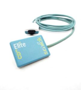
Schick CDR Elite Sensor is an easy-to-use design and a robust also combines outstanding image quality hard-wearing construction.
Schick CDR Elite Sensor combines outstanding image quality, a design that is quite easy to use, a proper construction that provides an excellent intraoral radiography experience, which is truly elite. It gives images for bold bone trabeculation, bright and clean DEJ, crisp lamina dura to meet the needs of every clinician in diagnosing.
The Schick CDR Elite Sensor embraces the technology of Schick’s removable cable and incorporates it in all its three sensor sizes, hence delivering convenience and simplicity of cable replacement to every clinician.
Its smart remote system is developed to allow accuracy and simplicity in in-office diagnosis. Schick elite sensor is a massive improvement in everything and is highly ranked in the industry. It integrates easily with CDR DICOM imaging software and Eaglesoft imaging.
It is equipped with enhanced features, high resolution, a sharpness that produces superb images for clinicians. This sensor is built to last for a long time, giving the best service to the clinicians who use it. With a warranty, this system is fully installed and serviced by very well-qualified Schick engineers.
4. Planmeca Prosensor HD ($2,500.00)
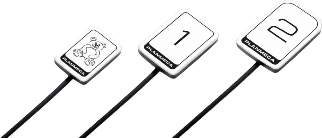
Planmeca prosensor HD intraoral sensor offers a very unique combination of high image quality, usability, and patient-centered design. With a resolution of more than 20lp/mm, it provides HD image quality. The planmeca prosensor HD has a fiber-optic layer that captures low noise, sharp and high contrast images.
It is available in three sizes. The sensor with edges that are round in design makes the procedure to be comfortable for patients while producing high-quality images in seconds. It is connected using either Ethernet or a USB port. The sensor has a magnetic connector that can be easily attached by hand. Its control box is equipped with a coded LED light that provides an instant visual of the imaging procedure.
The planmeca prosensor HD is designed to endure, its cable is reinforced and has two wires, and on top of that, there is a five-year warranty. It has layers that are designed to give perfect results. With a shock protector, ceramic LTCC circuit board, durable cables, indeed it is designed to last.
Planmeca prosensor HD has a wide dynamic range, a sealed housing, fully compatible with Mac and window OS, a plug then play USB version, can be integrated into the planmeca proX x-ray unit, and a planmeca Romexis software that has the most versatile tools. Indeed it is another dental x-ray sensor that truly has simplified dental health.
5. Apex dental sensors ($3,499.30)
If you have high standards for your dental equipment, then selecting the Apex dental sensors can help you. It features a high-resolution CMOS Sensor that can offer you crystal clear images. It can capture high resolutions images at 25 theoretical lines per millimeter.
It has a thin and elegant dental sensor design that is made for optimum comfort for your aspiring patients. The product also represents an excellent balance of function and form.
This sensor is designed with rounded corners. It helps to ensure easy placing for the comfort of the patient. It features easy-to-use and powerful imaging software for producing an exact diagnosis. It is also effortless to disinfect because it is 100% dustproof and waterproof. It is hermetically sealed and comes with an IP67 rating. The sensor can be immersed by sterilizing liquid without worrying that the sensor may get damaged.
The Apex dental sensor is unique because it can work with different imaging software. It can support TWAIN devices which allow communication between digital imaging devices and software. It also has excellent flexibility and durability. It passes through rigorous testing and designs for its maximum performance. It also comes with silicon padding and shock-resistant cases for protection from bites, falls, and other damage.
6. Dexis platinum sensor ($1,575.00)
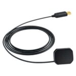
Beveled corners, around and smooth casing, and an angled dome, the dexis platinum sensor maximizes the patient comfort while making precise placement easy. Having a direct USB connectivity, its concept is portability, ease, and with Pure image technology, the quality you get of the images is remarkable.
The Dexis platinum sensor gives visible and detailed images. It has a cesium iodide scintillator that converts the x-ray beam into visible light, and through micro-columnar structures, it guides it to the sensor using the fiber optic. It brings detailed images via its CMOS sensor that maximizes the active is used by pixels.
The sensor prevents x-ray backscattering by having a protective shield that is fixed to the rear housing interior wall. The Dexis software and hardware work harmoniously to balance radiation variances hence guiding against over and underexposure.
Its imaging makes it possible to see subtleties, which are crucial in diagnosis, patient communication, and collaboration. Hugely detailed images from the dexis platinum sensor have made the digital x-ray easy for the detection of defects.
7. Vatech Ezsensor HD ($3,119.20)
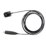
EzSensor HD provides the necessary high-resolution images for accurate diagnosis With a theoretical resolution of 33.78 lp/mm. New noise and artifact reduction image processing provides clear and consistent images, making the EzSensor HD one of the easiest to use in the market today.
The Vatech ezsensor is slim, with rounded corners for maximum comfort. The vatech ezsensor has a massive range of exposure settings, and it captures consistent images that can easily be diagnosed. The sensor is highly sensitive, capturing diagnosable images under various conditions, including when using old x-ray sources. The patients even then get lower radiation.
Its high contrast makes it possible to have a look at the insides’ different opacity in detail. It makes it easy to even distinguish interproximal carries in the image. The imaging of vatech is based on CMOS APS technology, which provides high resolution with more transparent images helping in diagnosis and treatment.
Its exterior is made from high-strength aluminum giving it protection. The cable is flexible to protect the sensor from damage. You can quickly and easily switch between seven contrast filters to get the best clear diagnostic image. It is directly connected to your computer with a USB, and you do not need a control box, hence making transportation easy and can be utilized in many operatories.
8. Gendex GXS-700 ($1,990.00)

Designed for smooth migration from a film, easy upgrading of a digital system, the Gendex gxs-700 is the 8th generation of Gendex. It comes with excellent imaging, portability, and easy to use. With advanced CMOS sensor technology that gives high-quality images, it elevates diagnostic capabilities.
It provides over 20 visible line pairs per mm, working with a wide range of settings that produces quality images and with repeatable results.
The Gendex gxs-700 has two sensor sizes that are ergonomically designed to accommodate adults and children. Their corners are rounded and smooth for patient comfort and to fit the oral cavity anatomical shape. Its materials are durable for a longer life span.
It has a 2.0 high-speed USB connectivity that does not require any adapters, docking stations, or USB controllers. This helps the Gendex gxs-700 to deliver high-quality real-time images with amazing clarity. Its HD images help capture challenging areas like third molars and also long-rooted canines.
The Gendex gxs-700 can quickly and conveniently be carried to any place it is needed. It is fast in delivering images, thus providing immediate feedback. It has many eco-friendly features that its core purpose is to reduce radiation.
9. RVG 6100 ($2,200.00)
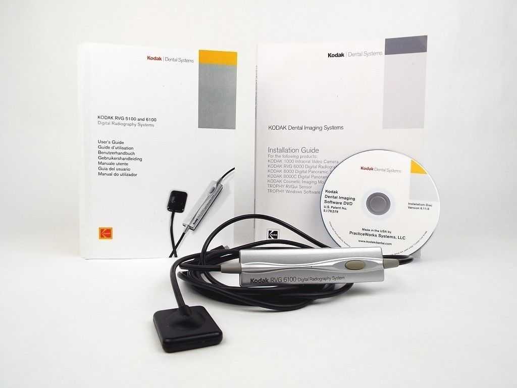
The RVG 6100 is cutting-edge technology in dental X-ray sensors. It is built to give an outstanding high-quality image that clinicians can use to diagnose and treat patients. It has a sharpness filter that emphasizes a radiological feature that in the transparent image, it can go unnoticed.
It has a highlight tool that is useful in detecting fractures and carries through the tool reinforcing contrast locally.
The RVG 6100 sensor has a fast 2.0 USB connection for quick delivery of crystal clear images for clinicians. It is designed to resist bites, shocks, and drops is designed to provide durability hence return of investment. It offers a higher radiation reduction than another RVG size one sensor.
The RVG 6100 digital imaging system has the highest resolution in the market today. With advanced technology in optical plate and scintillator, it allows lower radiation doses while providing high resolution for complex examination providing quick diagnoses for treatment.
Like many other sensors in the market, it has rounded edges to help make positioning easy while giving comfort to the patient.
10. RVG 6200 ($3,100.00)
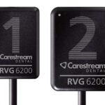
Carestream RVG 6200 features straightforward installation and integrates with most imaging and dental practice management software and makes sophisticated diagnostic imagery simple Carestream RVG 6200 features the CS Adapt module, a set of user-defined image processing tools that allow users to choose from either 40 pre-set image enhancement filters or define up to four favorite settings of their own, resulting in a customized comfort zone for every appointment. Don’t adapt to technology—let technology adapt to you.
RVG 6200 System is also one of the fastest digital imaging sensors on the market—displaying images in less than two seconds. A great solution for multi-chair practices, the sensor is designed to streamline your workflow and ensure easy image capture, analysis, and sharing. With such superb abilities, it’s not surprising to learn that many renowned oral healthcare professionals consider the RVG 6200 System the best intraoral radiographic system on the market.
Conclusion
Dental Digital imaging has changed dental x-ray sensors bringing the best in the industry. Looking at the above ten best digital imaging sensors, it is true the digital imaging has gone a milestone in producing high-tech sensors that deliver high-quality images with easy-to-use settings to give the best diagnosis.
They have reduced exposure to radiation for patients while increasing their comfort and easy positioning through their designs. They produce images fast, and some are portable, growing the number of patients that can be reached. They have well-crafted software that can be installed on computers for easy diagnosis.
Their durability, eco-friendliness, fast delivery of diagnosis put these sensors on a high level in digital imaging. They are affordable and consume less power.
These dental x-ray sensors have improved the dental department, and any clinicians should go for this amazing technology to get the best diagnosis in dental health.


No Comments